

Trigger Finger
What is trigger finger?
Stenosing tenosynovitis of the A1 pulley, also known as “trigger finger” results from the flexor tendons getting trapped under the sling that prevents the tendons from bow stringing on the palm of the hand.

Patients usually complain of the finger getting stuck in flexion (towards the palm), often requiring the use of force from the other hand to open. They may have a bump in the palm that can be painful to touch. (6,7)
How is it diagnosed?
It can be diagnosed on clinical examination, however, a dynamic diagnostic ultrasound with visualization of thickened pulley system, and tendons getting entrapped can confirm the diagnosis.
Common findings on diagnostic ultrasound include thickening of the A1 pulley, accessory blood vessels within the tendon sheath (neovessels), tendon enlargement/nodularity, tenosynovitis with or without hyperemia on Doppler, and abnormal tendon motion during dynamic flexion and extension of the finger with or without triggering. (6-13)
While occasionally post-traumatic, stenosing tenosynovitis at the A1 pulley typically develops insidiously and without a definable event. Common risk factors for development of stenosing tenosynovitis include diabetes, rheumatoid arthritis, prior carpal tunnel release, and gout. (14)
How is it treated?
Conservatively this can be treated with bracing, occupational therapy and watchful waiting. Corticosteroid injections have been shown to be beneficial in the treatment of trigger finger pulley (15-18). Ultrasound guided injections are more accurate than PG injection and are effective in reducing symptoms at 3-year follow-up (15). Injection with hyaluronic acid has also been demonstrated to reduce triggering at 3-month follow-up (19). Hyaluronic acid is not traditionally covered by insurances for this purpose and is expensive. For patients who have failed ultrasound guided steroid injection, traditional open release and percutaneous in office ultrasound guided release are treatment options.
What is ultrasound guided trigger finger release/injection?
The use of ultrasound guidance to perform release of the A1 pulley for the treatment of trigger finger, using needles or specialized cutting devices has been recently described with good results (20-28).
This involves the use of direct ultrasound guidance to guide a specialized needle to the pulley and cut the pulley. Research has shown that this approach has a decreased complication rate to open release and can safely and effectively be performed in the office with excellent results (20-28).
Following local numbing with lidocaine, using ultrasound guidance a specialized needle is introduced through a small poke hole in the palm, nothing that needs to be sutured. The whole procedure takes about 5-10 minutes from start to finish and the patient can immediately resume usual activities!
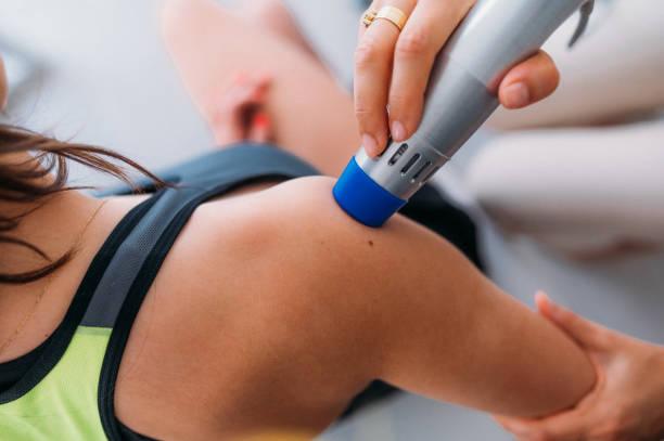
In house guided trigger release on a patient via USG.
How does this compare to the traditional open procedure?
It has been shown to have a lower complication rate than traditional open trigger finger release with less scarring and less need for post-operative pain medications (27).
There is ongoing research in this field and upcoming level 1 study being performed by Douglas Hoffman, MD in Minnesota, directly comparing the traditional open approach to the ultrasound guided approach. This information will be updated once published.
What are the risks/complications?
The risks of any procedure involving a break in the skin include, infection, neurovascular or tendinous soft tissue injury and failure to provide relief. Ultrasound makes risk of iatrogenic injury to neurovascular structures and anatomic variants lower as the anatomic is viewed throughout the procedure. According to research and my anecdotal experience this risk is lower than that of the traditional open approach (27). In the literature and my ongoing patient reported outcomes, there is a 10-15 % chance of persistent figure swelling that can last up to 3 months and require further injections to help resolve. This is compared to the open procedure which has up to a 43% chance of this occurring (27, 29-31).
What is the recovery like?
Anecdotally, my patients typically are sore the day of the procedure but do not require pain medications. They can immediately resume usual activities with care being taken not to soak the area for 24 hours.
Most patient on patient reported outcomes (Quick DASH), report rapid (4-14 day) return to activity pain free and immediate resolution of triggering (31).
Reference Articles
See the full list of Dr. Pourcho's reference publications on this topic
Research articles
Minimally Invasive Ultrasound-Guided CarpalTunnel Release: Preliminary Clinical Results
Henning PT, Yang L, Awan T, Lueders D, Pourcho AM.J Ultrasound Med. 2018Nov;37(11):2699-2706. doi: 10.1002/jum.14618. Epub 2018 Apr 2.PMID: 29608024
Sonographic Changes After Ultrasound-Guided Release of theTransverse Carpal Ligament: A Case Report.
Latzka EW, Henning PT, Pourcho AM.PM R. 2018Oct;10(10):1125-1129. doi: 10.1016/j.pmrj.2018.02.018. Epub 2018 Mar 6.PMID: 29518589
Wise A., Pourcho AM, P.T. Henning, Latzka EW. Evidence for ultrasound-guided carpal tunnel release- Review – Archives of PM R, 2020


Book an Appointment
We're Here to Help You
Ready to take the next step in restoring your physical health? Contact us today!